Your lungs hold secrets that a simple breathing test can unlock. When your doctor hands you a spirometry report filled with abbreviations like FEV1, FVC, and mysterious percentages, it might feel like trying to decipher a foreign language. Yet these numbers tell a powerful story about your respiratory health – one that could reveal hidden conditions or confirm that your lungs are functioning perfectly.
Spirometry remains the gold standard for measuring lung function, helping diagnose conditions from asthma to COPD. Understanding what those raw numbers mean empowers you to take control of your respiratory health and have meaningful conversations with your healthcare provider.
Understanding the Basic Spirometry Parameters
The foundation of spirometry interpretation rests on three key measurements that paint a comprehensive picture of your lung function:
FVC (Forced Vital Capacity) measures the total amount of air you can forcefully exhale after taking the deepest breath possible. Think of it as your lungs’ maximum storage capacity. Normal values typically range from 80% to 120% of the predicted value based on your age, height, sex, and ethnicity.
FEV1 (Forced Expiratory Volume in 1 second) captures how much air you can blow out in the first second of a forceful exhalation. This measurement proves particularly valuable because many lung diseases affect the speed at which you can empty your lungs.
FEV1/FVC Ratio represents the percentage of your total lung capacity that you can exhale in one second. This ratio serves as a crucial diagnostic tool – a value below 70% often indicates airflow obstruction, while normal values typically exceed 75-80%.
Reading Your Spirometry Report: A Step-by-Step Guide
When you receive your spirometry report, you’ll notice two columns that matter most: your actual values and the predicted values. The predicted values come from population studies and represent what someone of your demographics should theoretically achieve.
| Parameter | Your Result | Predicted Value | % Predicted | Interpretation |
|---|---|---|---|---|
| FVC | 3.5 L | 4.2 L | 83% | Normal (>80%) |
| FEV1 | 2.8 L | 3.4 L | 82% | Normal (>80%) |
| FEV1/FVC | 80% | 81% | 99% | Normal (>70%) |
The % predicted column reveals how your lungs compare to the expected normal range. Values above 80% for FVC and FEV1 generally indicate normal lung function. However, interpreting these numbers requires considering the complete clinical picture, including your symptoms and medical history.
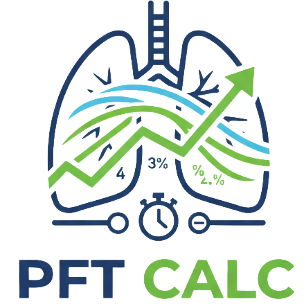
For accurate interpretation of your spirometry values, you can use the PFT Calculator to quickly determine your predicted values and understand where your results fall within the normal range.
Recognizing Patterns: Obstructive vs. Restrictive Disease
Spirometry results typically reveal one of three patterns that guide diagnosis:
Obstructive Pattern appears when airways narrow or become blocked, making it harder to exhale quickly. You’ll see:
- Reduced FEV1 (often below 80% predicted)
- Normal or reduced FVC
- Decreased FEV1/FVC ratio (below 70%)
- Common in asthma, COPD, and chronic bronchitis
Restrictive Pattern occurs when the lungs cannot fully expand, limiting the total amount of air they can hold:
- Reduced FVC (below 80% predicted)
- Proportionally reduced FEV1
- Normal or increased FEV1/FVC ratio
- Seen in pulmonary fibrosis, chest wall deformities, or neuromuscular diseases
Mixed Pattern combines features of both obstructive and restrictive disease, making interpretation more complex and often requiring additional testing.
The American Thoracic Society provides comprehensive guidelines at their official website for healthcare providers interpreting these patterns.
Advanced Parameters and What They Reveal
Beyond the basic measurements, spirometry reports often include additional parameters that provide deeper insights:
FEF 25-75% (Forced Expiratory Flow) measures airflow during the middle portion of exhalation. This sensitive indicator can detect early small airway disease even when FEV1 and FVC appear normal. Values below 65% of predicted may suggest developing airway obstruction.
PEF (Peak Expiratory Flow) captures the fastest speed you can exhale. While effort-dependent, significant reductions often indicate large airway obstruction or poor respiratory muscle strength.
FEV6 (Forced Expiratory Volume in 6 seconds) serves as an alternative to FVC in some settings, particularly useful for patients who struggle with prolonged exhalation.
The Role of Bronchodilator Response Testing
Many spirometry tests include a bronchodilator response assessment, where you repeat the test after inhaling medication that opens airways. This comparison reveals crucial diagnostic information:
A significant bronchodilator response means:
- FEV1 increases by at least 12% AND 200 mL
- FVC increases by at least 12% AND 200 mL
This reversibility strongly suggests asthma rather than COPD, though some COPD patients also show improvement. The absence of reversibility doesn’t rule out asthma, as inflammation levels fluctuate.
Quality Indicators: Ensuring Accurate Results
Not all spirometry tests provide reliable data. Quality indicators on your report ensure the results accurately reflect your lung function:
Acceptability Criteria include:
- Good start to the test (no hesitation or false starts)
- No coughing during the first second
- Smooth, continuous exhalation
- Maximum effort throughout
- Adequate exhalation time (at least 6 seconds)
Reproducibility requires at least three acceptable maneuvers with the two best FVC and FEV1 values within 150 mL of each other. Reports showing poor reproducibility may need repeating for accurate diagnosis.
Special Considerations for Different Populations
Interpreting spirometry results requires adjusting for individual factors:
Age-related changes naturally reduce lung function over time. After age 25-30, FEV1 typically declines by 20-30 mL per year in healthy individuals. What seems abnormal at 30 might be perfectly normal at 70.
Ethnicity affects lung volumes, with established correction factors for different populations. African Americans typically have lung volumes 10-15% lower than Caucasians of the same height and age, while Asian populations may have 5-10% lower values.
Body habitus influences results significantly. Obesity can restrict lung expansion, while very tall or short individuals may fall outside standard prediction equations.
The CDC provides detailed reference values at their NIOSH Spirometry page that account for these demographic factors.
From Numbers to Diagnosis: Clinical Integration
Spirometry results never stand alone in making a diagnosis. Physicians integrate these numbers with:
Clinical symptoms such as shortness of breath, cough, wheezing, or chest tightness provide context for interpreting borderline results. Someone with normal spirometry but significant symptoms may need additional testing.
Medical history including smoking, occupational exposures, family history, and previous respiratory infections shapes the interpretation. A 20-pack-year smoking history makes COPD more likely than asthma when seeing an obstructive pattern.
Physical examination findings like wheezing, decreased breath sounds, or chest wall abnormalities correlate with spirometry patterns and guide further evaluation.